AI Precision Oncology: Matching Tumors to Therapies
How multi-omic, imaging, and pathology fusion drives tumor profiling and therapy selection — and why data integration, not algorithms, is the hard part.

A tumor is not one data type. It is a genome, a transcriptome, a stained slide, a CT volume, a stack of pathology reports, and a treatment history — and the question every oncologist actually asks is narrow: given all of that, which therapy is most likely to work for this patient. For two decades the answer came from looking at each data type in isolation. The change underway in 2026 is that the most useful signal increasingly comes from fusing them, and the engineering problem is integration far more than it is modeling.
This is worth stating plainly because the marketing gets it backwards. The accuracy of any single classifier matters less than whether the genomic report, the slide, and the radiology read can be joined to the same patient, in the same encounter, in time for a treatment decision. That is a Data Platforms problem before it is an AI problem.
What “profiling a tumor” actually means now#
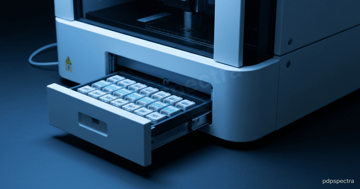
The baseline is comprehensive genomic profiling. Foundation Medicine’s FoundationOne CDx is an FDA-approved tissue-based assay that sequences 324 genes plus genomic signatures and carries companion-diagnostic claims tied to a long list of targeted therapies. Tempus recently received FDA approval for a tumor-only indication on its xT CDx platform, a 648-gene next-generation sequencing test that reports microsatellite instability status alongside its companion-diagnostic claims. These are regulated diagnostics, not research tools, and they define the floor: a structured, validated readout of what is broken in the tumor.
Genomics alone, though, leaves signal on the table. The same mutation behaves differently depending on expression, tumor microenvironment, and tissue morphology. That is why the field is moving toward fusion — combining DNA and RNA sequencing with the digitized pathology slide and, where available, radiology. An umbrella review of fusion strategies in AI cancer research lays out the three options cleanly: early fusion (concatenate raw or low-level features before modeling), late fusion (train per-modality models and combine their outputs), and hybrid approaches in between. Each has a different failure mode, and choosing among them is mostly an operational decision, not an accuracy contest.
Pathology is becoming a data source, not a verdict#
The digitized slide used to be the end of the line — a pathologist looked, signed, done. Now it is an input. Paige’s prostate-cancer detection software was the first AI product to receive FDA approval in digital pathology, and the research frontier has moved to foundation models trained on enormous slide archives. The Atlas pathology model, a collaboration between Mayo Clinic, Charité, and Aignostics, was trained on roughly 1.2 million whole-slide images across more than 490,000 cases, spanning dozens of tissue types and staining protocols. The point of a model like this is not to replace the read. It is to produce a dense, reusable representation of the slide that can be joined with genomic and clinical features.
That joining is where the value concentrates. Reported work shows AI systems integrating histopathology with genomic data to infer markers such as microsatellite instability directly from routine slides — a signal that would otherwise require a separate molecular test. Treat those figures as directional rather than guaranteed: they come from specific cohorts, and generalization across scanners, stains, and populations remains the open question. The responsible framing is decision-support — a second read that flags cases for molecular confirmation — not autonomous diagnosis.
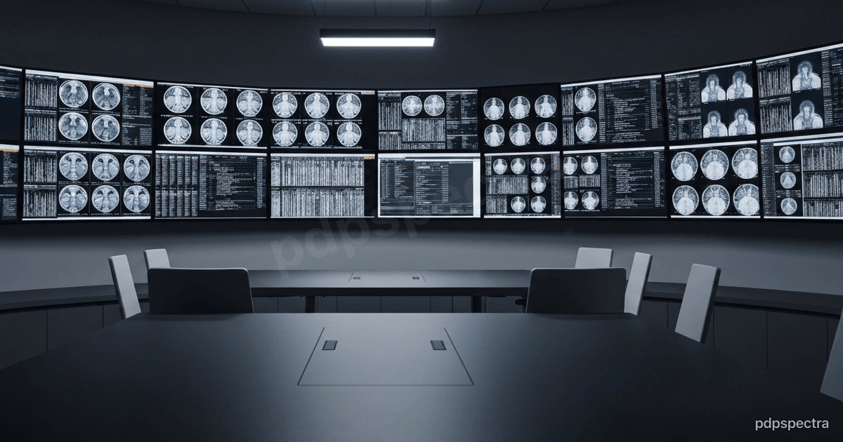
The molecular tumor board is the real integration point#
If you want to see where AI in oncology has to land, look at the molecular tumor board. This is the recurring meeting where oncologists, pathologists, radiologists, and geneticists reconcile a single patient’s genomic report, imaging, pathology, and history into a therapy recommendation. It is multidisciplinary precisely because no single modality is sufficient.
Recent work on AI in multidisciplinary tumor boards describes the pattern: electronic health records, imaging, pathology reports, and genomic data are processed through a stack of machine-learning, natural-language-processing, and large-language-model components that synthesize the inputs into diagnostic, prognostic, and therapeutic candidates. The board still decides. The system’s job is to make sure no actionable alteration and no relevant trial gets missed because the data lived in four different systems.
This is the part that consultancies actually get hired for. The model is rarely the bottleneck. The bottleneck is that the genomic report arrives as a PDF, the slide lives in a separate image-management system, the labs sit in the Hospital Management System, and the radiology lives in PACS — and nothing shares a stable patient and specimen identifier. AI implementation in oncology is, in practice, an exercise in entity resolution and pipeline reliability.
Therapy selection, grounded in evidence#
The most defensible AI in this space targets a specific, narrow decision rather than a general diagnosis. Tempus published a study in JCO Precision Oncology validating its PurIST algorithm for therapy selection in advanced pancreatic cancer, informing first-line chemotherapy choice from a molecular subtype. That is the right shape for clinical AI: one tumor type, one decision, validated against real-world outcomes, with a clear path into the standard of care.
Multi-omic integration extends this. A review of AI-driven multi-omics integration in precision oncology frames the goal as bridging the “data deluge” of genomics, transcriptomics, and imaging into something a clinician can act on. The honest assessment in that literature is that most multimodal models remain in research and validation, not routine clinical deployment. The regulatory reality is that anything informing treatment is a medical device, and the bar — analytical validation, clinical validation, post-market monitoring — is high for good reason.
Liquid biopsy widens the input set#
Tissue is not always available, and a tumor evolves after the biopsy is taken. Cell-free DNA assays address both gaps. FoundationOne Liquid CDx is an FDA-approved 324-gene assay that profiles circulating tumor DNA from a blood draw, which means a patient can be re-profiled longitudinally without another invasive procedure. For an integration layer this adds a temporal dimension: the same patient now has a sequence of molecular snapshots, and a resistance mutation that emerges on therapy shows up in the blood before it shows up anywhere else. A platform that treats genomic profiling as a one-time event misses this entirely. The data model has to be longitudinal, and the tumor board needs to see the trajectory, not just the latest result.
Where the engineering actually lives#
Strip away the modeling and the work that determines success looks like this.
Specimen-level identity. A patient has multiple specimens over time; a specimen has multiple blocks; a block has multiple slides and assays. Fusion is impossible without an identity model that tracks this hierarchy. Most failures trace back to a missing or ambiguous specimen key, not a weak classifier.
Modality availability is the norm, not the exception. Real patients are missing the RNA panel, or the slide never got scanned, or the imaging is from an outside facility. A fusion pipeline that only works when all modalities are present is a demo. Production systems have to degrade gracefully, which is one reason late-fusion designs — separate per-modality models whose outputs are combined — are easier to operate than early fusion: each component can be validated and monitored on its own and missing inputs do not break the whole.
Latency tied to the decision window. A profiling result that lands after the treatment decision is worthless. The pipeline’s service-level objective is set by the clinical calendar — the tumor board date — not by a benchmark. This is Operational Automation in the literal sense: turning a multi-week manual reconciliation into a reliable, scheduled process.
The data backbone. Imaging, labs, orders, and reports almost always originate in the Hospital Management System. If that system cannot emit clean, identified events for genomics and pathology to attach to, no amount of model sophistication recovers the loss. Get the backbone right and the AI layer becomes tractable; get it wrong and you are doing manual chart review with extra steps.
The honest position#
AI precision oncology in 2026 is real, regulated, and useful — within bounds. The validated wins are narrow and specific: a companion diagnostic that flags eligible patients, a subtype classifier that informs one chemotherapy choice, a pathology model that surfaces cases for molecular confirmation. The hype is the implication that a single multimodal model autonomously selects therapy. It does not, and the regulatory framework will not allow it to for the foreseeable future. The clinician and the tumor board remain the decision-makers, and that is correct.
For anyone building in this space, the durable advantage is not the model — those are increasingly commoditized and often open. It is the Data Platforms work underneath: clean specimen identity, reliable multimodal pipelines, and an integration layer that gets the right evidence in front of the board on time. That is unglamorous, and it is the whole game.
Building the data backbone for multimodal oncology? We integrate genomics, pathology, imaging, and EHR into pipelines clinicians can actually use. Talk to our engineering team.