AI in Digital Pathology in 2026: Vendors, Slides, and Clinical Use
Digital pathology AI matured in 2026 — PathAI, Paige, Tempus, Roche Navify. Whole-slide imaging, breast and prostate cancer detection, pharma partnerships, and the lab integration story.

A breast biopsy slide is digitized on a Philips IntelliSite Ultra Fast Scanner at 06:40 and lands in the pathology lab queue as a multi-gigapixel whole-slide image. Before the pathologist signs in, Paige Prostate (or the Paige Breast counterpart) has run a tumor-detection model across the slide, flagged a candidate invasive ductal carcinoma region, and pre-populated a draft synoptic report. The pathologist confirms the diagnosis, edits the staging fields, and signs out the case before the morning multi-disciplinary conference. Five years ago this was a slide on a microscope and a manually typed report.
Digital pathology is the second-most-mature radiology-adjacent AI domain in 2026. It lagged radiology by roughly a decade because the slide-digitization step is operationally heavy — scanners are expensive, slide volumes are immense, and the files are gigapixel-scale. The economics finally crossed over for breast, prostate, and colorectal cancer workflows, and the vendor landscape consolidated meaningfully over 2024–2026.
The vendors actually shipping in production#
PathAI sits at the centre of the platform conversation — they shifted from a pure model-developer to a full lab-integration platform with PathOS, ingesting whole-slide images from Leica Aperio, Philips, Hamamatsu, and 3DHistech scanners and orchestrating both their own and partner models. Strong pharma-services revenue funds the FDA clearance pipeline.
Paige.AI — acquired by Tempus in 2024 — was the first vendor to win FDA clearance for an AI-based pathology product (Paige Prostate, 2021). Under Tempus, the prostate, breast, and lymph node detection products integrate into the Tempus oncology data platform, which is the strategic logic of the acquisition.
Tempus itself anchors a broader oncology data play — sequencing, imaging, and clinical data — with pathology AI now as a workflow surface. Their reach into community oncology practices is meaningful and gives the pathology models a route into clinical workflow that pure-play vendors struggle to match.
Roche Navify Digital Pathology is the OEM-bundled stack from the diagnostics giant, tightly integrated with Ventana stainers, immunohistochemistry workflows, and the Roche slide-management infrastructure. Adoption follows the Roche IHC install base.
Indica Labs HALO AP is the lab-information-system-adjacent platform — strong in research and pharma workflows, with a growing clinical footprint. Their viewer is the de facto standard in many academic pathology departments.
Visiopharm, Aiforia, Owkin, Mindpeak, Ibex Medical Analytics, Proscia — each with niche strengths around breast, prostate, gastric, dermatopathology, or lab-platform integration.
Whole-slide scanner OEMs — Leica Biosystems (Aperio GT 450), Philips IntelliSite, Hamamatsu NanoZoomer, 3DHistech Pannoramic, Roche Ventana DP — are the upstream substrate that every AI vendor must integrate with.
What the technical stack looks like#
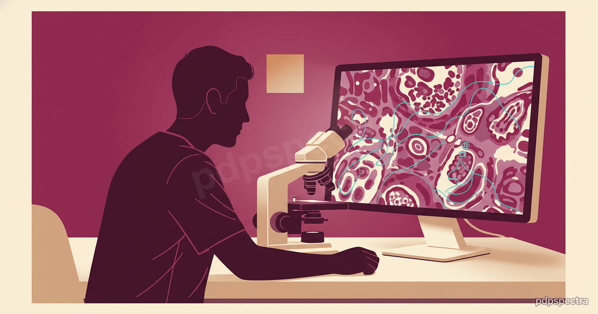
A whole-slide image is a pyramidal tiled file in formats like SVS, MRXS, NDPI, or DICOM WSI, often 1–10 gigapixels and several gigabytes per slide. AI models — typically convolutional networks or vision transformers — operate on tile-level patches and aggregate predictions at slide level. Training datasets are pathologist-annotated regions across thousands of cases, with the better vendors stratifying by lab, scanner type, stain protocol, and patient demographics to manage generalization risk.
The inference pipeline is non-trivial. Slides are written from the scanner to a storage layer (often an object store with a DICOM WSI gateway), tiled and pre-processed, sent through one or more models, and the predictions are written back as either an overlay heatmap, structured annotations, or pre-populated report fields. Latency targets are looser than radiology — pathologists sign out cases on a daily cycle, not in real time — but throughput matters because a busy academic lab may digitize fifty thousand slides per month.
Integration with the laboratory information system (LIS) — Sunquest, Epic Beaker, Cerner CoPathPlus, NovoPath, Sysmex CaseWorks — is where deployments succeed or fail. The model output must reach the pathologist inside the LIS or the case-management viewer, not in a separate browser tab, or adoption decays within weeks.
The breast, prostate, and colorectal clinical use cases#
The strongest clinical evidence sits in three cancers.
Prostate. Paige Prostate and the Ibex Galen Prostate product both have published reader-study data showing meaningful improvements in detection sensitivity and turnaround time. The use case — flag the cancer-containing core, draft Gleason grading — is well-bounded and aligns with how the pathologist actually works.
Breast. AI-assisted detection of invasive carcinoma, ductal carcinoma in situ, and lymph-node metastases. Mindpeak, Paige, and PathAI ship breast products. Lymph-node micrometastasis detection is a particularly strong use case because the manual screening burden is high and the AI sensitivity advantage compounds.
Colorectal. Polyp histology classification, tumor budding, microsatellite-instability prediction from H&E directly — the last one is genuinely interesting because it reduces downstream molecular test burden. Multiple vendors ship colorectal AI.
Other use cases — dermatopathology (Proscia, DermAI), hematopathology, renal pathology — are at earlier maturity.
Pharma partnerships are the quiet revenue engine#
A meaningful share of pathology AI revenue comes not from hospital labs but from pharma. PathAI’s biopharma services, Tempus’s oncology data partnerships, Owkin’s clinical-trial deals — pharma companies pay handsomely for AI-assisted analysis of trial-arm biopsies, biomarker discovery, and companion-diagnostic development. This revenue funds the clinical-side product development and explains why the pure-play startups can sustain the multi-year FDA clearance cycle.
The structure of these deals matured through 2023–2025. Early-stage discovery work — image-based biomarker discovery, immune-cell-infiltrate quantification, spatial-biology analyses — is paid on a fee-for-service or platform-access basis. Companion-diagnostic co-development, where the AI vendor builds the regulated diagnostic that supports a pharma asset’s label, carries milestones plus royalties on the approved drug. The latter category is small in count but disproportionate in long-term value, and several pathology AI vendors structured their corporate strategy around landing one or two such deals per year.
Real-world-evidence services are the third revenue line. Tempus and a handful of platform vendors use their pathology-plus-clinical data assets to support post-market drug-effectiveness studies, payer-mandated outcome studies, and regulatory-grade evidence generation.
Regulatory state in 2026#
Paige Prostate Detect was the first FDA-cleared AI pathology product (2021); the cleared list has grown steadily since. The CE-IVDR transition in Europe forced many pathology AI vendors through a heavier conformity assessment than the previous IVDD regime, and a handful of products were temporarily withdrawn from the EU market during the transition. The FDA’s Predetermined Change Control Plan framework is now used routinely for pathology AI clearances, allowing model updates within pre-specified bounds.
CAP (College of American Pathologists) accreditation requirements increasingly reference AI validation — labs deploying clinical AI need documented validation on their own scanner-stain-population combination, not just the vendor’s clearance package.
Where pdpspectra fits#
We help pathology departments and reference labs design the integration layer between scanners, image storage, AI vendors, and the LIS — slide routing, model orchestration, result write-back into the pathologist’s reporting workflow, and the validation logging required for accreditation. See our data engineering practice for the gigapixel-storage and pipeline work.
Related reading#
- AI in clinical trials in 2026
- Healthcare AI playbook: pilot to production
- Voice AI in clinical documentation
If you are evaluating digital pathology AI for a hospital lab, reference lab, or pharma-services partner and want a vendor-neutral read on what actually fits your slide volume and case mix, reach out.